|
Studies on the effects of ionization on bacterial aerosols in a burns and plastic surgery unit
By PAAVO MAKELA* Helsinki University Central Hospital, Helsinki JUHANI OJAJARVI Department of Public Health Science, University of Helsinki, Helsinki GUNNAR GRAEFFE ANm MATTI LEHTIMAKI Department of Physics, Tampere University of Technology, Tampere
(Received 1 August 1978)
SUMMARY
The effect of the ionization of the air on the decay of bacterial aerosols was studied in a Burns and Plastic Surgery Unit. Ions were generated by free corona needles. The air content of bacteria measured by settle plates was found to be smaller during the ionization period than during the controls period. The number of individual phage typed Staph. Aureus strains was especially found to be lower during ionization. The opposite potential increased the disappearance of bacteria from the air. The size of skin particles carrying bacteria is not optimum, but the results obtained show that the ionization may have applications in controlling airborne infection.
INTRODUCTION
Air contains electrically charged air ions produced in nature usually by radiation such as radioactivity, cosmic radiation or ultraviolet light. After ionization small air ions are formed by clustering of 10-20 molecules. Air ions can be generated with electric corona, discharge. These ions have either positive or negative charge depending on the polarity of the corona, voltage. It has been suggested that air ions formed by negative corona current exert favoUrable metabolic effects on the human being (Krueger & Reed, 1976; Gualtierotti, Kornblueh & Sirtori, 1968). The growth of the colonies of some microorganisms is altered and the decay of aerosol is faster (Krueger & Reed, 1976). Owing to ionization, the air ions move towards the opposite potential represented by walls, ceilings, floors, etc. The speed of the movement depends on the size of the air ions and their charge (Lehtimäki & Graeffe, 1976). The purpose of this study has been to examine the possibilities of decreasing bacterial contamination of the air by ionization.
* Requests for reprints: Paavo Mäkelä, Helsinki University Central Hospital, Paasikivenkatu 4, 00250 Helsinki 25, Finland.
0022-1724/79/0097-1978 $01.00 C) 1979 Cambridge University Press
200
METHODS AND DESIGN OF THE STUDY
The study was conducted in the Burns and Plastic Surgery Unit of Helsinki University Central Hospital. One part of the ward is reserved for bums. It consists of four rooms with necessary bath, service, storage and nurses' rooms. The ventilation of the ward is natural with no mechanical aids. During the winter the relative humidity of the ward was 13-34 % and the temperature varied from 20 to 24°C. The only mechanically ventilated room was a single bedroom where two burned patients studied were nursed, one at a time, on a Munter's ventilated air cushion. This causes one air change in the room every hour. In this room the relative humidity of the air was exceptionally low, from 12 to 20
The production of ions
In the first phase of the study ions were produced by a negative ion generator at - 5 or - 8 kV. Four free corona needles were hung at the height of 2 m, one on each side of the patient's bed and two at the other end of the room equidistant from walls and from each other. During the patient experiments three metal plates with a diameter of 9 cm to collect bacteria were situated vertically below the window and had opposite charges to that of the corona needles. In the second phase of the study all patient rooms and the corridor were equipped with corona needles.
Bacteriological methods
Air samples were collected by using settle plates with I h exposure time. The nutrient medium was ordinary blood agar. The agar was not earthed, as the number of colony counts was found to be consistent with or without earthing the media. Bacterial samples from metal plates were taken at the end of the study periods by contact plates (Hall & Hartnett, 1964) using nutrient agar with no additives. In preliminary experiments the collection time of settle plate samples was half an hour and the samples from metal plates were taken after the 2 h experiment. The bacterial samples were incubated at 37°C overnight and kept for another clay on the laboratory bench before identification and counting of bacterial colonies. The identification of Staph. Aureus was based on morphology, pigmentation of the colonies and tube coagulase test. The identification of other bacteria was based on gram-staining, colonial morphology and standard bacteriological techniques.
Patients 1. Three patients of the first phase of the study were nursed in single rooms. The first one was a paraplegic with decubital ulcers on major trochanteric areas. Staph. Aureus, phage type 85, was repeatedly isolated from his lesions. The second patient had burns of 30 % of the skin surface. He was nursed on a Munter's air cushion. Staph. Aureus, phage type 84, was cultured from his burns. The corona charge in this experiment was - 5 kV.
201
Table 1. Bacterial contamination of the air (settle plates) and on the earthed metal surface (contact plates) with or without the ionization of the air (charge - 5 k V) and/or charge ( + 5 k V) of the metal surface (The settle plates were changed every half an hour and the time of experiment was 2 h. The current was switched on at the beginning of the test.) Mean colony counts/2 h Mean colony Ionization Metal on the counts/2h of the air surface settle plates contact plate
No Uncharged 6.8 12.6 Yes Uncharged 7.6 17.4 Yes Charged 5.5 59.2 No Charged 8.4 29.8 No Uncharged 17.3 12.8
The third patient had bums of 40 % of the skin surface area. Bacterial cultures from her burns yielded an untypable Staph. Aureus, phage type NT. She was also nursed on Munter's air cushion. The corona charge in the experiments with this patient was - 8 kV. In these three experiments tests were carried out on alternate days. The ion generators were switched on at 9 p.m. on the preceding day and off at 9 p.m. on the test day. Air samples were collected at first from 7 a.m. until 7 p.m. and in later experiments from 8 a.m. to 4 p.m. Five settle plates were used simultaneously in a room. 2. In the second phase the air of single rooms was ionized as in the first phase of the study, and additional free corona needles were installed in other rooms and corridor to maintain equal ionization of the air in the whole unit. The ion generators were kept on every other week. The study was conducted during a 5-week period starting with a control week. Each week period started on Friday night. Bacteriological samples were taken as before on Tuesday and Thursday from 8 a.m. until 5 p.m. During four study weeks the patients and their number in the ward varied. Only one patient with 40 % burns was uninterruptedly nursed in the same room for 11 weeks. His Staph. Aureus was untypable.
RESULTS
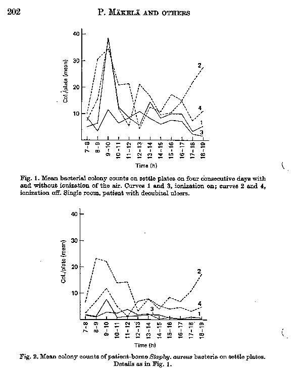
The preliminary experiments revealed that the earthed metal surface yielded more bacteria when the air was ionized (Table 1). The numbers of bacterial colonies on the metal plate substantially increased when the plate was charged to + 5 kV. The charged metal plate collected bacteria also without ionization of the air, but the number of colonies was lower. 1. Hourly variations in mean bacterial colony counts on settle plates in the single room of the first patient are shown in Fig. 1. Most of the colonies were Staph. epidermidis strains. High colony counts were caused by bed-making, dressing of wounds and other activities. With the exception of one high count, 13-2
the bacterial colony counts of the air during ionization were on a lower level than those during control days. The difference between mean colony counts during control and ionization days is statistically significant (t-test, P < 0.01). The difference between Staph. Aureus (phage type 85) colony counts on settle plates during control and ionization days was even more pronounced (Fig. 2). The results of the study of the second patient were consistent with those of the JIM one. Raising the corona charge to - 8 kV did not improve the results (the third
203
Table 2. Mean bacterial colony counts on 1 h settle plates during ionization and control weeks (The means are calculated from all the samples of the same day. The charge of the corona needle was - 5 kV.) Ionization Ionization Day Control week week Control week week Tuesday, 13.1± 0.64 5.9± 0.30 8.8± 0.47 4.4 ±0.23 (N = 300) (N= 24 1) (N= 250) (N= 249) Thursday 10.5± 0.59 4.7± 0.26 9.7± 0.57 3.3± 0.23 (N = 300) (N= 265) (N= 250) (N= 180)
Table 3. Isolation frequency and mean colony counts of Staph. Aureus on 1 h settle plates during ionization and control weeks (The means are calculated from all the samples of the same day. The charge of the corona needle was - 5 kV.) Ionization Ionization Control week week Control week week Frequency 109/600= 45/506= 168/500= 9/429= of Staph. Aureus 18.2% 8.9% 33.6% 2.1% positive plates
Mean colony Tuesday 5.4 2.4 4.8 2.1 counts/ settle plates ( N= 300 (N= 241) (N = 250) (N = 249) Thursday 2.9 4.7 8.2 0 (N= 300) (N= 265) (N= 250) (N = 180)
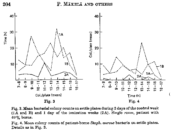
patient). The bacterial cultures from metal plates revealed three to five times higher colony counts during the ionization period than during control period The mean number of Staph. Aureus colonies from the metal plates was also two to five times higher during the ionization period than during control period. 2. In the experiments of weekly test periods the mean daily colony counts can settle plates during control weeks were twice as high as those during ionization weeks (Table 2). The difference is statistically highly significant (t-test, P < 0-001). Although the number of Staph. Aureus bacteria in the air was low, it was found that they -were less often isolated during the ionization period (Table 3). The metal plate cultures showed more bacteria when the ion generator was on. During ionization. the number and the frequency of Staph. Aureus bacteria were also higher. During the test week the total counts on metal plates were about ten times higher than those during control weeks. One patient stayed for 1 1/2 weeks in the same room. The mean number of total bacterial colony counts averaged 13-5 during the two control days (control week). The corresponding figure for the following ionization weeks was 3-8. From settle
plates Wing the control week Staph. Aureus bacteria were almost invariably isolated whereas these bacteria were rarely cultured during the ionization week (Figs. 3 and 4). During ionization total colony counts on the metal plates were high compared with those obtained with the ion generator off . The counts during the control days ranged from two to ten colonies/contact plate whereas during the ionization days the corresponding Figures were from 10 to 67. During the ionization period Staph. Aureus counts on the metal plates were high.
DISCUSSION
In studies in, a closed space, the half life of the aerosol particles has been found to correlate with the size of the particles and the charge of the corona current (Lehtimäki & Graeffe, 1976). Small, 0.01 mm size particles in the aerosol move fastest when the air is ionized. Phillips, Harris & Jones (1964) have noticed in their studies with Serratia Marcescens that negative ionization of the air leads to faster decay of the microbial aerosols than positive ionization. The number of microbes in the air decreases with the accentuated disappearance of the dust from the air. The ionization has been found to inhibit the air transmission of Newcastle virus bearing particles in experiments with chickens (Estola, Mikeli & Hovi, 1979). The size of virus-bearing droplets from the airways is considerably smaller than that of microcolonies on skin scales. Most human-borne bacteria are carried in the air on epithelial cells with a mean diameter of 20 mm (Noble & Somerville,
205
1974Y It would be expected that their disappearance from the air is slower than that of small droplets. Thus the ionization may not be as effective with bacterial aerosols originating from the skin. The electromagnetic field is weaker with corona needle ionization than with tunnel ionization and electrofilters . However, the continuous wider reaching effect results in ionization without additional turbulence caused by fans and ozone formation of the higher voltages. In this study we have measured the total bacterial air contamination mainly comprising Staph. epidermidis bacteria originating from human beings. The air contamination from individual patients was represented by phage typed Staph. Aureus strains. The bacteria-collecting effect of the metal plates shows that the opposite potential increases the disappearance of the aerosol. This was noticed by increased numbers of viable bacterial colonies on the positively charged metal surface. During both 24 h and 7-day ionization periods the colony counts on settle plates were low and occasionally no identified Staph Aureus were found in patient rooms. However, charged metal plates showed high bacterial counts with Staph. Aureus strains originating from the patient. The sedimentation of bacteria correlated with the air content of bacterial microcolonies during the ionization. The air measurements were done by filtration techniques using gelatine filters (Koller & Rotter, 1974). The number of bacterial colonies was consistent with or without earthing the culture media of the sedimentation plates. Gravity therefore determines the direction of fallout for sedimenting particles and the settle plate technique is thus a valid indicator of the air content of bacteria. As numerous cultures were needed in this kind of long lasting study, the settle plate method was the best alternative. It is known that the microbial contamination of the air in burns units is high (Hambraeus, 1973). In single rooms where isolated patients were nursed the ionization experiments of 24 h periods with - 5 kV showed lower sedimenting bacterial counts during ionization on two repeated occasions. The total colony counts represent contamination due to the staff and the patients. Phage typed Staph. Aureus strains in the air indicate shedding by individual patients. Although the she of bacteria-carrying epithelial cells is large, the number of Staph.. Aureus bearing particles was significantly decreased by ionization. A partial explanation may be that the shedding of the particles is inhibited by their immediate charging when separated from the patient. This causes the fixing of the particles back to their origin. The effect of ionization was apparent even with low values of relative humidity. Although measurements done in a closed space without air movement were in favour of voltages higher than - 5 kV our measurements with - 8 kV did not yield better results (Lehtimäki & Graeffe, 1976). However, by increasing the intensity of the electromagnetic field with the opposite potential the particles were more effectively drawn towards vertical surfaces. In the burns unit studied the patient rooms have no airlocks to the hall. The isolation facilities are poor and the mixing of the air is evident. The number of patients, the variation of individual patients and the activity of the nursing staff
206
varied during the 4-week period. Nevertheless, the difference in total colony counts of the air was significant between control and ionization periods. The number of Staph. Aureus shed by patients in the presented cases was also lower during the ionization. The continuous shedding of Staph. Aureus by the patients was verified by their presence on the positively charged metal plates. Ionization experiments with animal respiratory disease caused by Newcastle disease virus (Estola et al. 1979) suggest that the contamination of the air by droplets that carry bacteria such as Mycobacterium tuberculosis, Mycoplasma pneumoniae, etc. may be prevented by ionization of the air. Further studies on this are needed- As an energy saving method with low running expenses ionization of the air may prove to be an alternative to increased air ventilation and filtration.
We thank Mrs Tioni Sorsa, R.N., Maria Ratia, R.N. and Beatrice Uotila, laboratory technician, for the technical assistance. We are grateful for the cooperation of all the staff of the Bums and Plastic Surgery Unit. Ion generators were supplied by Ilmasti Oy, Helsinki, Finland.
REFERENCES
BARR, P. 0., BIRK, G., LILJEDAHL S. 0. & PLANTIN, L. 0. (1907). Treatment of burns with warm dry air. Lancet ii, 1276. ESTOLA, T., MÄKELÄ, P. & Hovi, T. (1979). The effect of air ionization on the air-borne transmission of experimental Newcastle disease virus infections in chickens. Journal of Hygiene 83, 59. GUALTIEROTTI, R., KORNBLUEH, I. H. & SIRTORIi, L. (1968) The influence of ionization on endocrine glands . In Bioclimatology, Biometeorology and Aeroionotherapy, Milan, Kaarlo Erba Foundation. HALL, L. B. & HARNETT, M. J. (19641. Measurement of the bacterial contamination on surfaces K hospitals. Public Health Reports, Washington 79, 102 1. HAMBRAEUS, A. (1973). Studies on transmission of Staphylococcus Aureus in an isolation ward for burned patients. Journal of Hygiene 71, 171. KOLLER W. & ROTTER M. (1974). Weiters untersuchungen Uber die Eignung von Gelatinfiltern zur Sammlung von Luftkeimen. Zentralblatt far Bakteriologie, Parasitenkunde, Infektionskrankheiten und Hygiene, Abt. I, Orig. B 159, 546. KRUEGER, P. & REED E(1976). Biological impact of small air ions. Science, N.Y. 193, 1209. LEHTIMÄKI M. & GRAEFFE G. (1976). The effect of the ionization of air on aerosols in closed spaces. Proceedings of the 3rd International Symposium on Contamination Control, Copenhagen, 2, 370-82. NOBLE, W. C. & Somerville D. A. (1974). Microbiology of Human Skin. London: Sanders. PHILLIPS G., HARRis G. J. & JONES M.V. (1964) Effect of air ions on bacterial aerosols. international Journal of Biometeorology 8, 27-37.
|
Copyright © Info-Systel S.A. 1999,2000,2001 Contact : ContactOrions@info-systel.com